The Mouse Hospital
The Mouse Hospital is a core facility within our laboratory led by Steve Sastra and Carmine Palermo. It is designed to recreate all aspects of a clinical trial while using mouse models. Mice genetically engineered to develop pancreatic cancer are bred centrally and then screened by high resolution ultrasound for the development of spontaneous pancreatic tumors. Tumor volumes are quantified via 3D ultrasound and, upon reaching a predetermined size, the tumor-bearing mice are randomized into treatment groups.
The advantage of this core structure is that each week a cohort of roughly synchronized tumors are simultaneously enrolled in a single experiment; this is in contrast to having several smaller project-specific colonies that generate just a few mice per week. The core approach allows researchers to focus on other experiments when not enrolling mice, providing increased efficiency of resources, both in terms of material and time. This structure demands a highly cooperative team of researchers and a dedicated staff scientist to manage the core mouse colony and carry out the pre-enrollment screening,
The Mouse Hospital plays a key role in our laboratory's translational research and focus on translational precision medicine. We are able to do this by collaborating with oncologists and surgeons at CUIMC to develop PDX models. The Mouse Hospital is uniquely organized to support the development of PDX models because it is designed to model the clinic. When our lab receives patient derived tumors it will be divided in several ways depending on the quality and quantity of the sample. Because we are a translational research lab, priority is given to the development of in vivo PDX models. The rest of the pieces are either sent for RNA sequencing, tumor banking, single cell assays, and/or the development of organoids and explants. It is important to note that we stay in compliance with the Institutional Review Board (IRB) when collecting patient samples, as well as the Institutional Animal Care and Use Committee when studying and caring for our animals. It is our priority to stay in compliance with these committees to continue generating ethical data.
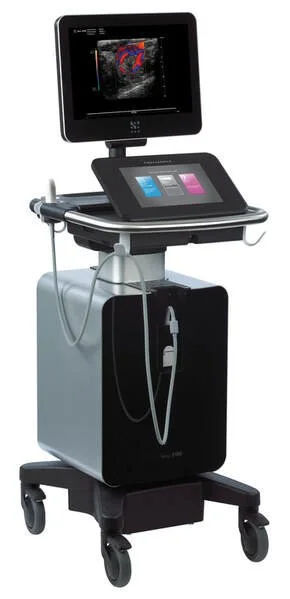
The Mouse Hospital core is located in a dedicated procedure room within the barrier facility in the ICRC building. It includes the Vevo 3100 ultrasound instrument, small animal anesthesia apparatus, and areas/equipment for drug preparation/administration and for surgical preparation, operation, and recovery.
Imaging
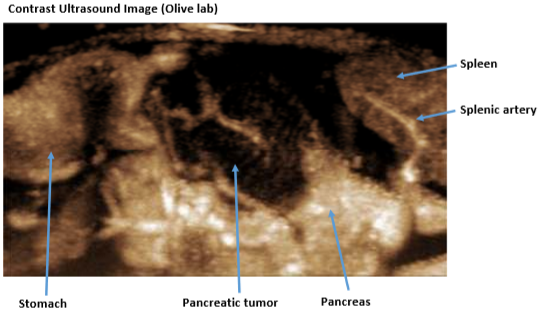
Imaging technologies are a powerful means to answer fundamental biological questions and advance translational research.
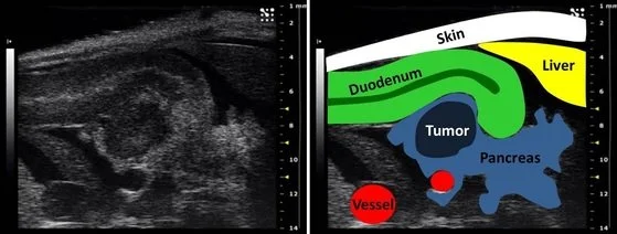
Our group utilizes high resolution ultrasound in order to detect, monitor, and quantify pancreatic tumors in mice. Using the upgraded VisualSonical Vevo 3100 high resolution ultrasound instrument, we can detect nascent tumors and premalignant lesions as small as half a millimeter in diameter.
On the right is an ultrasound image of the head of the pancreas of a mouse with a 4mm diameter tumor. On the right is a color-coded map of the anatomical features in this image. Key: white= skin; green= proximal duodenum; yellow- liver; red= large blood vessels; light blue= normal pancreas; dark blue= pancreatic tumor.
OPTIC’s manager, Chris Damoci, extensively highlighted of the broad potential of imaging in our research and other cancer research.